
Mammography
Mammography can detect cancer at its earliest stages, significantly improving healing rates.
Mammography is the radiological technique dedicated to the study of the breast. It is the basic and essential imaging method for diagnosing breast disease and the only one recognized as a screening technique for breast cancer, allowing early detection. It is also the only technique that has been shown to reduce breast cancer mortality rates.
It is particularly sensitive in the detection of microcalcifications, which may constitute an early sign of breast cancer and are difficult or impossible to detect using other imaging techniques (ultrasound, magnetic resonance imaging).
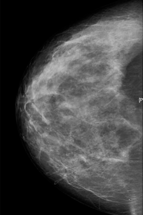
Mammogram view
When a mammogram is indicated
At our clinic, we perform an annual mammogram starting at the age of 40.
If the patient has an increased risk of breast cancer based on certain personal or family medical histories, their primary care physician may recommend starting the test before this age. It is important to note that mammography is not recommended before the age of 30.
Both the radiation risk and the economic cost are considered sufficiently low to justify its widespread use.
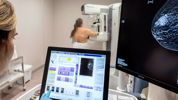
How a mammogram is performed
Our clinic is equipped with digital technology for performing mammograms and is among the most technologically advanced in Spain.
The diagnostic protocol for breast pathology should begin with anamnesis (medical history taking) and physical examination (inspection and palpation), performed by the technician prior to the radiographic study. These will determine the application of the remaining diagnostic techniques.
Two views must be systematically obtained: craniocaudal and mediolateral oblique. These may be supplemented with additional views (localized compression, magnification, etc.) to better assess suspicious areas, at the discretion of the radiologist.
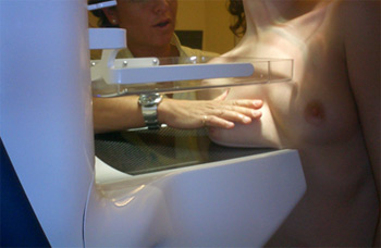
Performing a mammogram
Preparation
Mammography does not require any special preparation.
For a complete assessment of the mammographic study, it is essential that the woman bring any previous mammograms she may have at the time of the examination. This allows the radiologist to perform a comparative and evolutionary analysis of any possible findings.
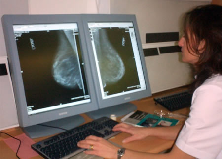
Digital mammography
After the test
After the mammogram, the radiologist may request a breast ultrasound if deemed appropriate, either due to high breast density or to evaluate a palpable nodule or a finding detected on the mammogram.
Mammography can also be used as a guide for pre-surgical localization of lesions or to guide needle biopsies using stereotactic techniques. However, whenever a lesion is visible on ultrasound, this will be the technique of choice, due to its speed and because it allows direct visualization of the lesion during the procedure.
Patient Care Information
Most health insurance providers cover this test with prior authorization. When you request an appointment, you will be informed about your insurance coverage and the procedures to follow.
At our clinic, you can receive the test report on the same day as the examination.


