
Breast ultrasound
Breast ultrasound is a technique that:
- Is essential, together with mammography, for the complete evaluation of patients with breast disease.
- Improves the specificity of mammography in the characterization of nodules.
- Is usually the imaging method of choice for guiding interventional procedures.
It is an examination that uses ultrasound waves (no ionizing radiation is used) to evaluate the internal structure of breast tissue. It is indicated for the assessment of masses or nodules detected during breast examination by palpation, or to further characterize abnormalities detected on mammography.
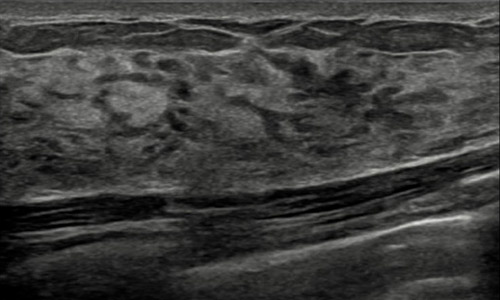
How breast tissue appears on an ultrasound
When a breast ultrasound is indicated
Ultrasound allows the differential evaluation of nodules that may be palpated by the gynaecologist during the clinical examination or that are visualized on mammography.
Breast ultrasound does not replace screening mammography, due to its inability to detect microcalcifications (calcium deposits that may sometimes indicate the presence of a small cancer). Only in the case of pregnant women and young women is it used as the initial diagnostic method instead of mammography.
Indications for breast ultrasound:
- It allows differentiation between a solid nodule and one with liquid content (cyst) and improves the characterization of solid nodules (as a complement to mammography).
- It is a diagnostic complement in patients with dense breast tissue.
- Evaluation of axillary lymph nodes in the context of a recently diagnosed breast carcinoma.
- As a guide for minimally invasive diagnostic procedures such as fine-needle aspiration or core needle biopsy.
- It is used to localize non-palpable lesions with wire localization prior to surgery and for clip placement. Both the wire and the clips serve as guides for the surgeon to ensure accurate removal, allowing for precise surgery.
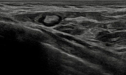
Normal axillary lymph node on ultrasound
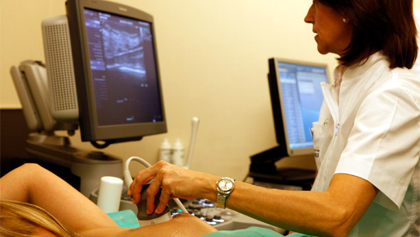
How a breast ultrasound is performed
The test is not uncomfortable and poses no risk to the patient.
During the ultrasound examination, the radiologist applies a water-based gel to both breasts, which allows better contact and transmission of the transducer over the skin, improving image quality.
The examination usually does not take long, generally no more than 10–20 minutes.
Preparation
The only preparation required is for the patient to uncover the area of the body to be examined —in this case, the breasts— and to lie calmly and relaxed on the examination table.
It is very important, if previous examinations were not performed at Dexeus, to bring any prior breast imaging studies.
Patient Care Information
Most health insurance providers cover this test with prior authorization. When you request an appointment, you will be informed about your insurance coverage and the procedures to follow.
At our clinic, you can receive the examination report on the same day as the test.


